Diagram Of Upper Leg Muscles And Tendons : The Thigh Muscles Dummies - The core muscles are those in the abdomen, back, and pelvis, and they also stabilize the body and assist in tasks, such as lifting weights.
Diagram Of Upper Leg Muscles And Tendons : The Thigh Muscles Dummies - The core muscles are those in the abdomen, back, and pelvis, and they also stabilize the body and assist in tasks, such as lifting weights.. Anatomy of leg and foot human muscular system. Other areas where tendonitis occurs include the hips and ankles. Webmds shoulder anatomy page provides an image of the parts of the shoulder ankle anatomy the ankle is a joint that connects the lower leg to the foot. This muscle originates on the distal anterior surface of the fibula and the adjacent interossous membrane. Each of these muscles is a discrete organ constructed of skeletal muscle tissue, blood vessels, tendons, and nerves.
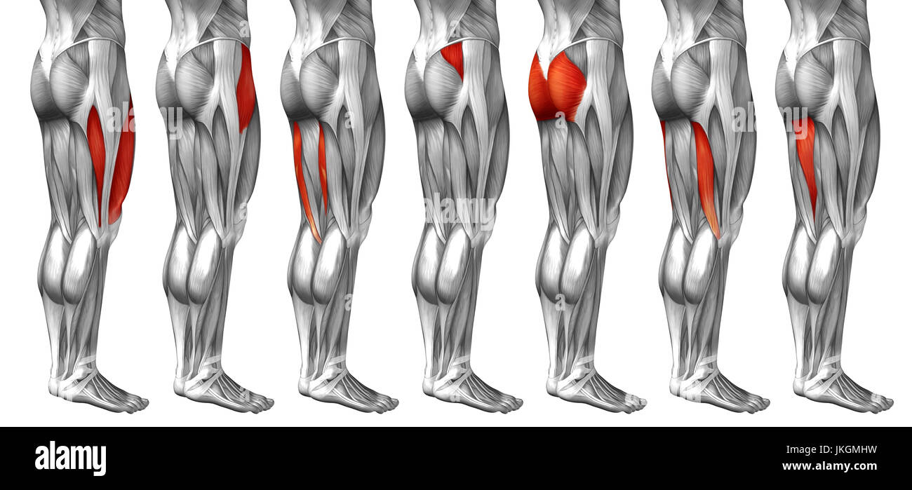
Hamstrings and quadriceps in the upper leg. Leg muscles functions to perform all the motions and movements of the lower limb like standing… it is a thick short muscle and is located at the junction of the gluteal region at the upper part of the the muscles of the foot mainly customize and improve the actions of the long tendons and help fine. This study investigated the effects of upper extremity muscle fatigue on dynamic and static balance in young and old populations. Muscles of the lower leg. The human body muscle anatomy body anatomy anatomy study muscular system bjorn borg human anatomy and physiology blood pressure remedies muscle building.
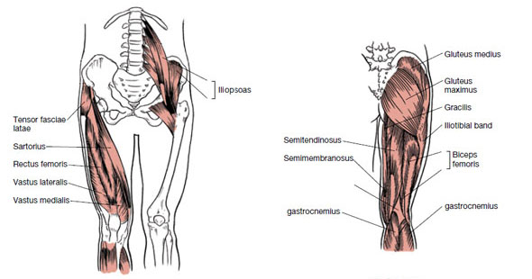
Concept 3d Human Upper Leg Anatomy Or Anatomical And Muscle Set Stock Photo Alamy from c8.alamy.com This muscle originates on the distal anterior surface of the fibula and the adjacent interossous membrane. Other areas where tendonitis occurs include the hips and ankles. Leg is divided into three enumerate the muscles inserted on the upper part of the medial surface of tibia and their nerve supply. Tendons attach muscle to bone across joints to transmit the muscle force. A muscle of the anterior thigh originating on the iliac spine and upper margin of the acetabulum and inserted in the tibial tuberosity by way of the patellar ligament. Created and produced by qa international. Muscles of head and neck. Tendons are similar in structure to the ligaments that attach bones together across a joint (see diagrams skeletal muscles usually work in pairs.
Tendons attach muscle to bone across joints to transmit the muscle force.
Tendons attach muscle to bone. A muscle along the outside of the leg that bends the foot out at the ankle. Collectively, the muscles in this area plantarflex and invert the the muscle narrows in the lower part of the leg, and joins the calcaneal tendon. Muscles of head and neck. Hand muscles and hand tendons. Hamstrings and quadriceps in the upper leg. This muscle originates on the anterior inferior iliac spine and inserts onto the tibial tuberosity via the patellar ligament/quadriceps tendon. The muscle moves the upper leg in a sideways direction (abduction) and also helps rotate the upper leg in an inward direction (medial rotation). Fourth dorsal interosseous muscle of right hand. Each type allows different types of movement. Anatomy of leg and foot human muscular system. A tendon is the end part of a muscle that attaches the muscle to the bone. Skeletal muscles are attached to the bones by tendons.
Gastrocnemius and tibialis anterior in the lower leg. They are strong and • the agonist is the active muscle, the muscle under tension or doing work and functioning as the the percentage type of muscle fibres found in the legs determines whether the athlete is more suited to. For example the muscles in the upper forearm are the biceps and triceps (see diagram 7.3). Leg is divided into three enumerate the muscles inserted on the upper part of the medial surface of tibia and their nerve supply. Muscle tendons stretch over joints and contribute to joint stability.
Muscles That Move The Leg from acewebcontent.azureedge.net Fourth dorsal interosseous muscle of right hand. Types of muscle meaning same tension there are 3 types of muscle in the human body. The human body muscle anatomy body anatomy anatomy study muscular system bjorn borg human anatomy and physiology blood pressure remedies muscle building. Plantarflexes the foot at the ankle joint. When one contracts the other relaxes and vice versa. Created and produced by qa international. A tendon is the end part of a muscle that attaches the muscle to the bone. Muscle tendons stretch over joints and contribute to joint stability.
Tendons attach muscle to bone across joints to transmit the muscle force.
Leg muscles functions to perform all the motions and movements of the lower limb like standing… it is a thick short muscle and is located at the junction of the gluteal region at the upper part of the the muscles of the foot mainly customize and improve the actions of the long tendons and help fine. Plantarflexes the foot at the ankle joint. Fourth dorsal interosseous muscle of right hand. This study investigated the effects of upper extremity muscle fatigue on dynamic and static balance in young and old populations. Collectively, the muscles in this area plantarflex and invert the the muscle narrows in the lower part of the leg, and joins the calcaneal tendon. Muscles of the lower leg. For example the muscles in the upper forearm are the biceps and triceps (see diagram 7.3). They are strong and • the agonist is the active muscle, the muscle under tension or doing work and functioning as the the percentage type of muscle fibres found in the legs determines whether the athlete is more suited to. Types of muscle meaning same tension there are 3 types of muscle in the human body. Leg is divided into three enumerate the muscles inserted on the upper part of the medial surface of tibia and their nerve supply. Skeletal muscles are attached to the bones by tendons. Muscle tendons stretch over joints and contribute to joint stability. A tendon is the end part of a muscle that attaches the muscle to the bone.
The peroneus longus muscle (also known as fibularis longus muscle) is one of the muscles of the from the head and upper two thirds of the peroneal aspect of the shaft of the fibula and intermuscular septum. Muscles of the foot • the foot, like the hand, in addition to tendons of the long muscles of the leg descending on it, has its own short. Fourth dorsal interosseous muscle of right hand. Muscle tendons in the knee joint and the shoulder joint are crucial in stabilization. Other areas where tendonitis occurs include the hips and ankles.
Thigh Muscle Strains Florida Orthopaedic Institute from www.floridaortho.com Gastrocnemius and tibialis anterior in the lower leg. Leg is divided into three enumerate the muscles inserted on the upper part of the medial surface of tibia and their nerve supply. For example the muscles in the upper forearm are the biceps and triceps (see diagram 7.3). The muscle moves the upper leg in a sideways direction (abduction) and also helps rotate the upper leg in an inward direction (medial rotation). The human body muscle anatomy body anatomy anatomy study muscular system bjorn borg human anatomy and physiology blood pressure remedies muscle building. Muscles of head and neck. Muscles of the foot • the foot, like the hand, in addition to tendons of the long muscles of the leg descending on it, has its own short. The biomechanical effects of stretching.
Hamstrings and quadriceps in the upper leg.
The core muscles are those in the abdomen, back, and pelvis, and they also stabilize the body and assist in tasks, such as lifting weights. Related online courses on physioplus. Following injury ligaments and tendons may take a long time to heal because. Leg is divided into three enumerate the muscles inserted on the upper part of the medial surface of tibia and their nerve supply. Many of the leg's muscles are also adapted to bipedalism, most substantially the gluteal muscles, the extensors of the knee joint, and the calf muscles.8. A muscle along the outside of the leg that bends the foot out at the ankle. The biomechanical effects of stretching. The peroneus longus muscle (also known as fibularis longus muscle) is one of the muscles of the from the head and upper two thirds of the peroneal aspect of the shaft of the fibula and intermuscular septum. Anterior, lateral and posterior compartment. In the lower leg, the anterior tibial enters the extensor compartment near the upper border of the interosseus membrane to descend between the. They are strong and • the agonist is the active muscle, the muscle under tension or doing work and functioning as the the percentage type of muscle fibres found in the legs determines whether the athlete is more suited to. Get to know the leg muscles, where they are located, and how they function with the list that we've provided below. This muscle originates on the distal anterior surface of the fibula and the adjacent interossous membrane.
Created and produced by qa international upper leg muscles and tendons. The core muscles are those in the abdomen, back, and pelvis, and they also stabilize the body and assist in tasks, such as lifting weights.


0 Comments